The Biology of the Goat
Development of the mammary gland in the goat
Birth At birth the doe already has a well defined outline of teats and gland cisterns.
Groups of cells, called sprouts have formed branches from the gland cistern.
These branches will become the ducts and alveoli in the mature udder.

Pre estrous
Between birth and puberty, before the doe first comes into heat, adipose (fatty)
and connective tissue are deposited in the mammary gland.
Sprouts have continued to grow and branch.

Estrous
During each estrous cycle, the ducts continue to grow and branch.
Estrogen is primarily responsible for the growth of the ducts.

After the end of each estrous cycle, there is some regression of duct growth.
The amount of regression is less than the amount of growth resulting in overall positive gain.
There is an increase in duct development of about 8% (in dairy cattle) with each estrous cycle.

Pregnancy
Most growth of the mammary gland occurs during pregnancy.
In the early stages of pregnancy, the ducts branch and grow.

During the middle and later stages of pregnancy, the milk-secreting alveoli form.
Progesterone acting with estrogen is responsible for the formation of alveoli.
Ducts continue to branch and grow.

In the last few weeks of pregnancy the alveoli begin secreting some milk products.
High levels of estrogen and progesterone inhibit lactation.
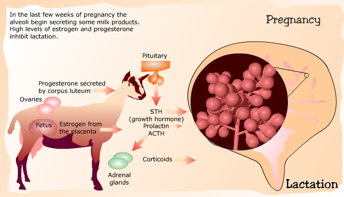
Lactation (initiation)
When the doe gives birth, the levels of estrogen and progesterone in the blood drop to
low levels which stimulate the production of prolactin by the pituitary gland.
Along with adrenal corticoids and growth hormone, prolactin is responsible
for the initiation of lactation.

Lactation Maintenance
Lactation is maintained by STH (growth hormone), thyroid stimulating hormone, and prolactin.
The sucking stimulus and milk removal are also essential to maintain lactation.

Involution
Involution (shrinkage) of milk secreting tissue naturally occurs sometime after the peak
of lactation causing reduced milk yield.

Milk secreting tissue regresses toward the cisterns.
Alveoli decrease in size and number, and the number of cells of each alveoli decrease.
Alveolar cell milk secreting activity decreases.
Later, whole lobules disintegrate leaving only branching ducts and connective tissue.

If the milking stimulus and milk removal is stopped, the increased pressure inside
the alveoli causes the cells to rupture and degenerate.
Milk is secreted into the connective tissue, and is broken down by the white blood cells.
Involution is much more abrupt.

The mammary gland tissue will regress almost to the same state as before pregnancy.
