The Biology of the Goat
The Anatomy of Lactation
The udder of the goat consists of two separate mammary glands.
Each half has one teat.

The medial suspensory ligament is made up of two sheets of elastic yellow tissue.
Each strong sheet covers the flat surface of the inside of each half of the udder.
The medial suspensory ligament connects to the pelvic symphysis supporting the udder
in a perfect balance.

Lateral suspensory ligaments have many layers which support the udder.
Deep layers surround the udder and pass into the gland.

Looking at one half of the udder in cross section shows the inside of the teat cistern
which is continuous with the gland cistern.
The teat and gland cisterns are also called the lactiferous sinus.

The teat ends in folds that form a star-shaped slit.
The folds are held closed by circular muscles which retains the milk and prevents dirt
and bacteria from entering the udder.

Blood is supplied by the mammary artery, and blood is drained by the mammary or the milk vein.

Lymph vessels remove fluid from around tissues.
Lymph fluid travels in only one direction, out of the udder, is filtered in the lymph node
and then leaves by a large lymph vessel.
Udder edema occurs when excessive lymph fluid accumulates under the skin of the udder.
The movement of lymph fluid is caused by pressure in the blood vessels,
by breathing, and muscle contraction.

Lymph vessels have valves that allow the lymph fluid to move in only one direction.

The tissue of the udder called the parenchyma consists of alveoli which look like bunches of grapes.
Cells lining the alveoli produce milk.

Each alveolus is surrounded by cells called basket cells which act like muscles.

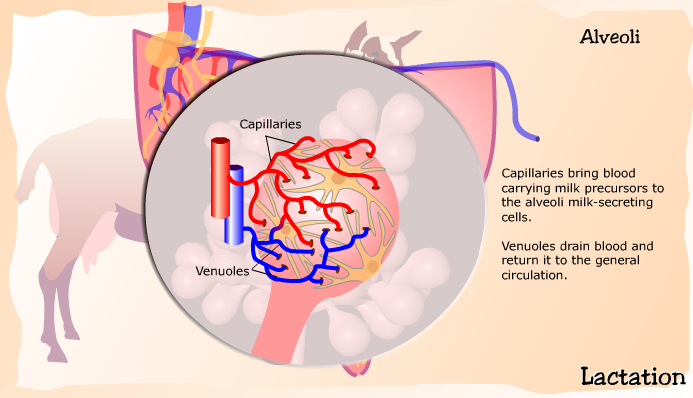
Capillaries bring blood carrying milk precursors to the alveoli milk-secreting cells.
Venuoles drain blood and return it to the general circulation.
Milk secreting cells line the inside of the alveolus.
When the alveolus is empty the cells are columnar.

As milk fills the inside (lumen) of the alveolus the cells stretch and become flattened.

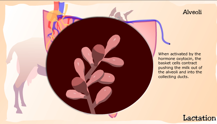
When activated by the hormone oxytocin, the basket cells contract pushing the milk
out of the alveoli and into the collecting ducts.
Flesh-colored secretory tissue is made up of alveoli that join a common duct.
White connective tissue surrounds bundles of alveoli forming a lobule.
A group of lobules are surrounded by connective tissue forming a lobe.
Lobes connect to larger ducts that lead to the milk cisterns.
