The Biology of the Goat
Cryptosporidium parvum

Oocysts seen at high power.
6 µm X 6 µm. (About 1/6 the size of Eimeria oocyst shown in background).
Floats high against the coverslip.
Life cycle
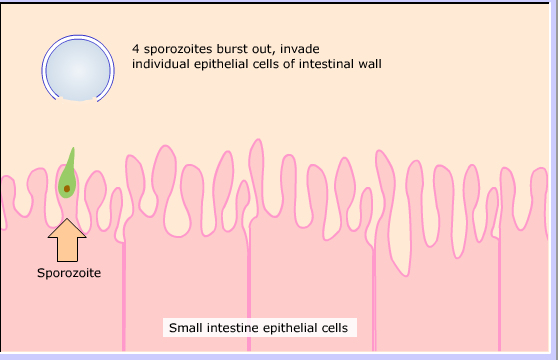
Oocyst enters small intestine.

4 sporozoites burst out, invade individual epithelial cells of intestinal wall
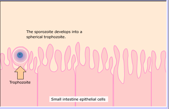
The sporozoite develops into a spherical trophozoite.
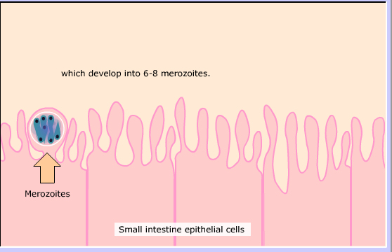
Asexual division produces 6-8 nuclei.

which develop into 6-8 merozoites.
Merozoites escape to invade new cells.

Can form more type I meronts

Or can form type II meronts.

Type I merozoites invade more cells (self infection)
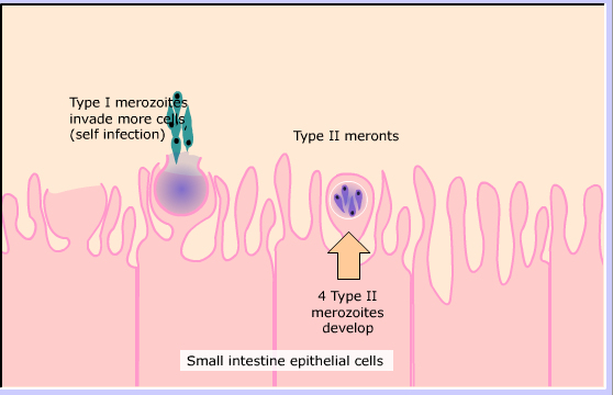
4 Type II merozoites develop
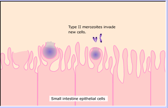
Type II merozoites invade new cells.

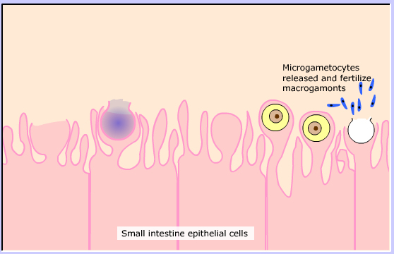
The merozoites differentiate into either male or female stages.

Microgametocyte (male) Divides into 16 microgametocytes Macrogamonts (female)

Microgametocytes released and fertilize macrogamonts
Zygotes formed
Meiosis occurs

Wall forms around oocyst containing four sporozoites
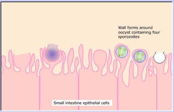
About 20% are thin-walled oocysts which reinfect more cells (second type of self infection).
Millions of thick-walled oocysts passed in the feces to infect another animal.

Clinical Signs
The parasite affects mostly very young animals of most species
causing severe or fatal diarrhea lasting several days or longer.
It can infect humans of all ages.
Other than supportive care, there is no effective treatment.
Exposure can result in full or partial immunity.