The Biology of the Goat
Parelaphostrongylus tenuis
Meningeal deer worm
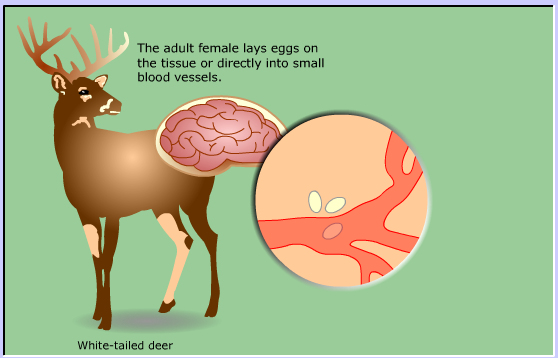
The adult brain worm lives in the connective tissue that surrounds the brain of white-tailed deer.
Meninges is the name for the tissue that surrounds the brain and spinal cord
explaining why another name for this parasite is the meningeal worm.

The adult female lays eggs on the tissue or directly into small blood vessels.
The eggs hatch and the larvae pass into the blood where they are carried to the lungs.
Eggs laid in the blood vessels hatch in lungs
The first stage larvae enter the respiratory tract and are coughed up then swallowed.
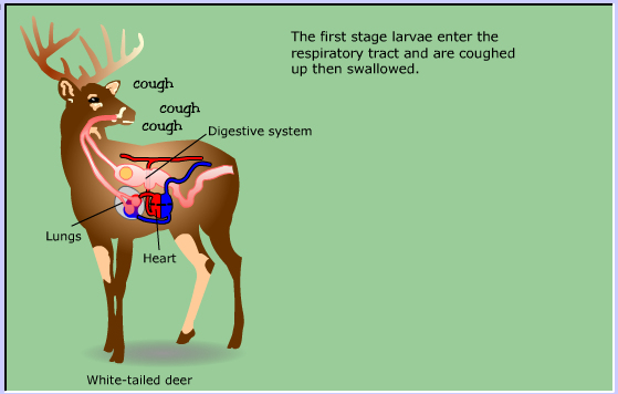
The larvae enter the digestive system and are passed out with the feces.
Land and water snails feast on the fecal pellets ingesting the larvae.
The snail is the intermediate host.

Inside the snail, the larvae grow through the 2nd and 3rd stages which takes about 3 to 4 weeks.

Deer accidently eat the snails while feeding.
The 3rd stage larva are released in the abomasum or small intestine,
then find their way to the spinal nerves and to the spinal cord.
This takes about 10 days.

The larvae mature in the gray matter of the spinal cord.
In about 1 month the mature adult migrates to the space around the spinal cord and travels to the brain.

It takes about 3 months from the time the deer is first infected until the adult brain worm
begins to lay eggs and repeat the cycle.
If a goat (or other herbivore) eats the snail the course is similar, but the larvae migrate randomly
through the nervous system causing severe neurological disease.
The goat is a dead end host.
The parasite does not reproduce in the goat.
Clinical signs
The deer rarely shows any symptoms of the infection.
Depending on where in the nervous system the worm migrates, goats and other herbivores show weakness, incoordination, gait abnormalities, lameness, partial or complete paralysis, dementia, stupor, coma, visual abnormalities, circling, falling or rolling, weakness, head tilt, altered head and neck position, seizures and death. Clinical signs usually begin in the hind limbs.